RAMONA
BLOG
Thank you! Your submission has been received!
Oops! Something went wrong while submitting the form.
Quantifying Cell Confluency at Scale: Parallel Plate Imaging with Automated Segmentation
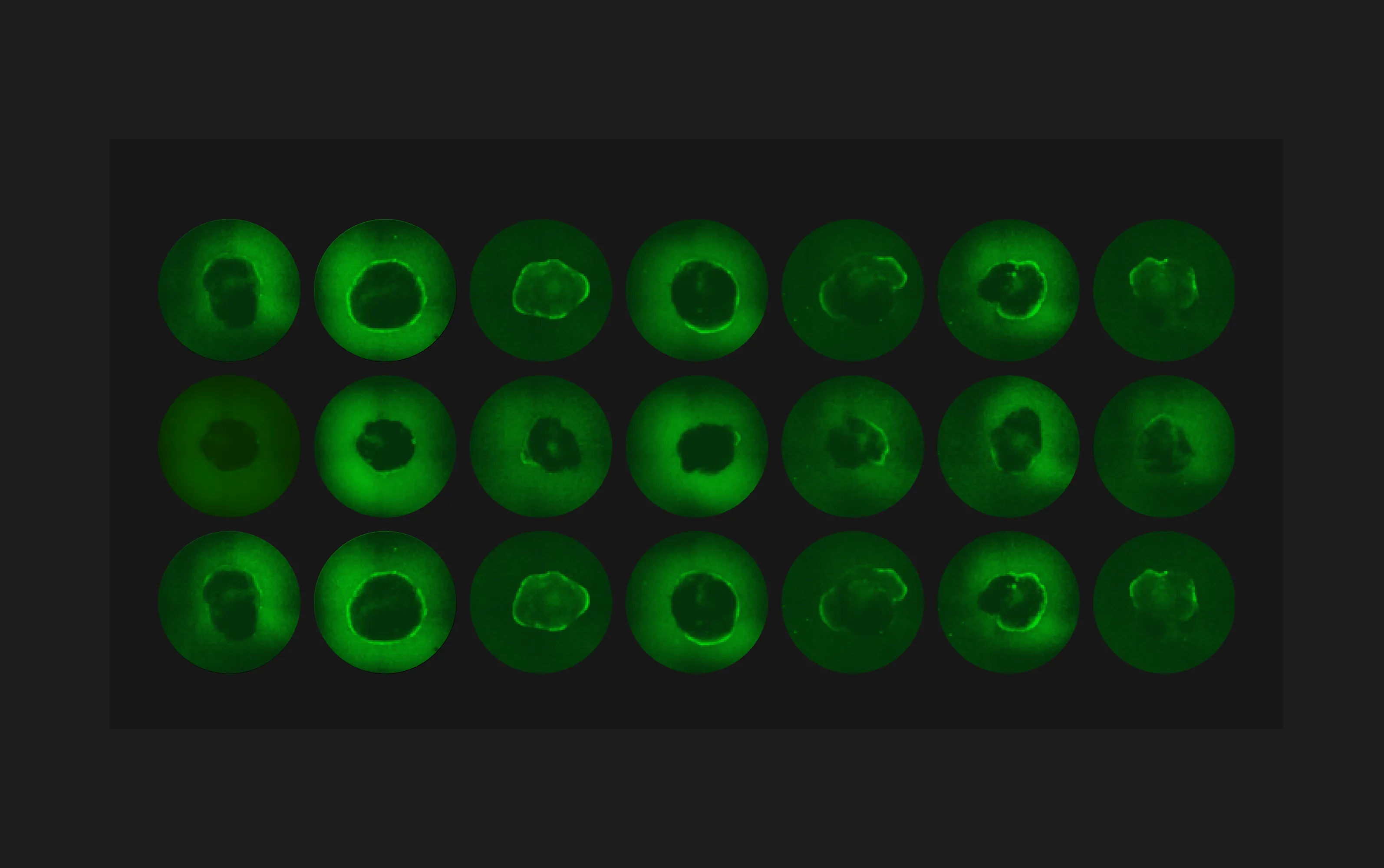
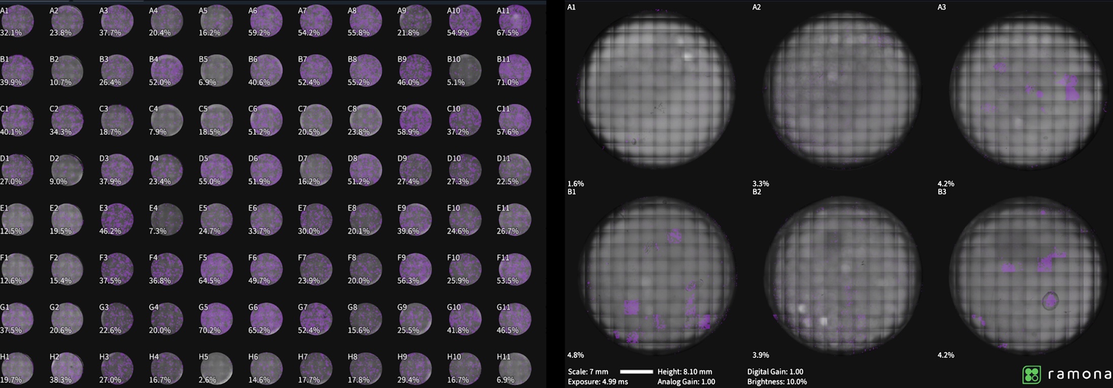
The Vireo enables automated cell confluency measurement across standard SBS plates (6, 12, 24, 96, or 384 wells) using parallel whole-plate imaging and full z-stack acquisition. Confluency is quantified as a percentage of well area for each well and exported as a CSV file with associated metadata. Example configurations include 4X imaging with 10 z-slices at 100 µm step size (30 seconds for a 96-well plate) and 10X imaging with 20 z-slices at 20–30 µm step size (2 minutes for a 96-well plate)..png)
.webp)
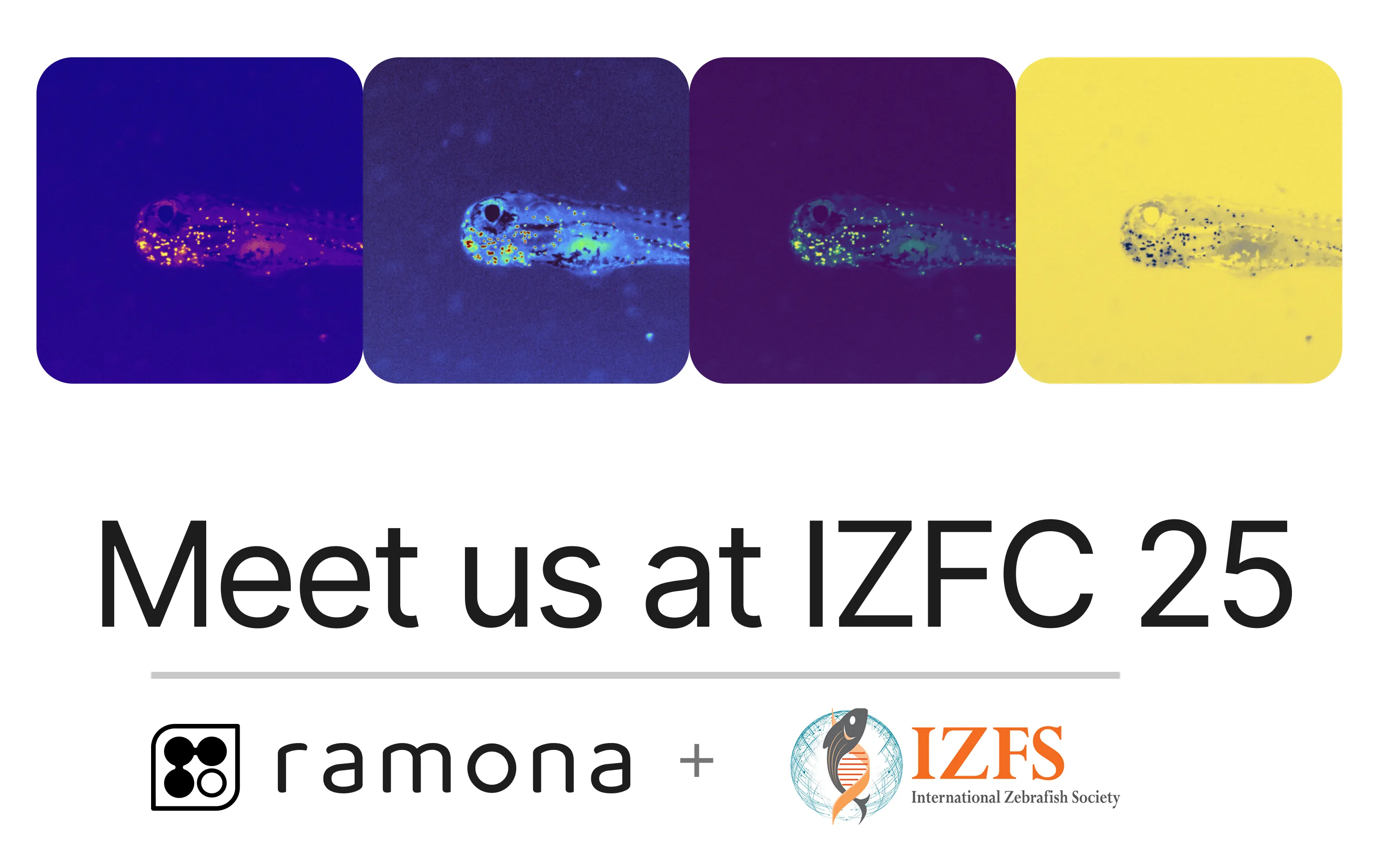
Scaling Functional Imaging with Parallelized Microscopy [WATCH NOW]
Dr. Franz Kuchling (Tufts University) and Dr. Patrick Fortuna (Harvard Medical School & Wyss Institute) reveal how Ramona’s parallel imaging technology is redefining functional biology, scaling real-time insights from living systems like Volvox and cardiac organoids through unprecedented speed and field of view..webp)


Ready to transform your research?
Book a call with one of our Ramona experts: