Expand
Your Field of View

Ramonaā€™s microscopy toolset frees researchers from the constraints of traditional imaging. Propel your research with Ramona.
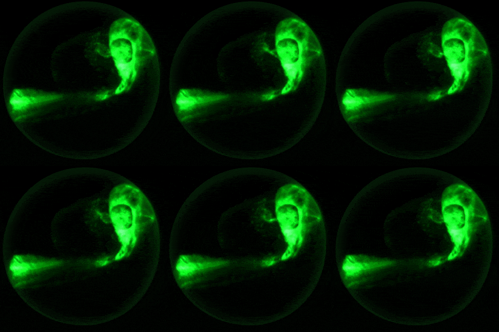
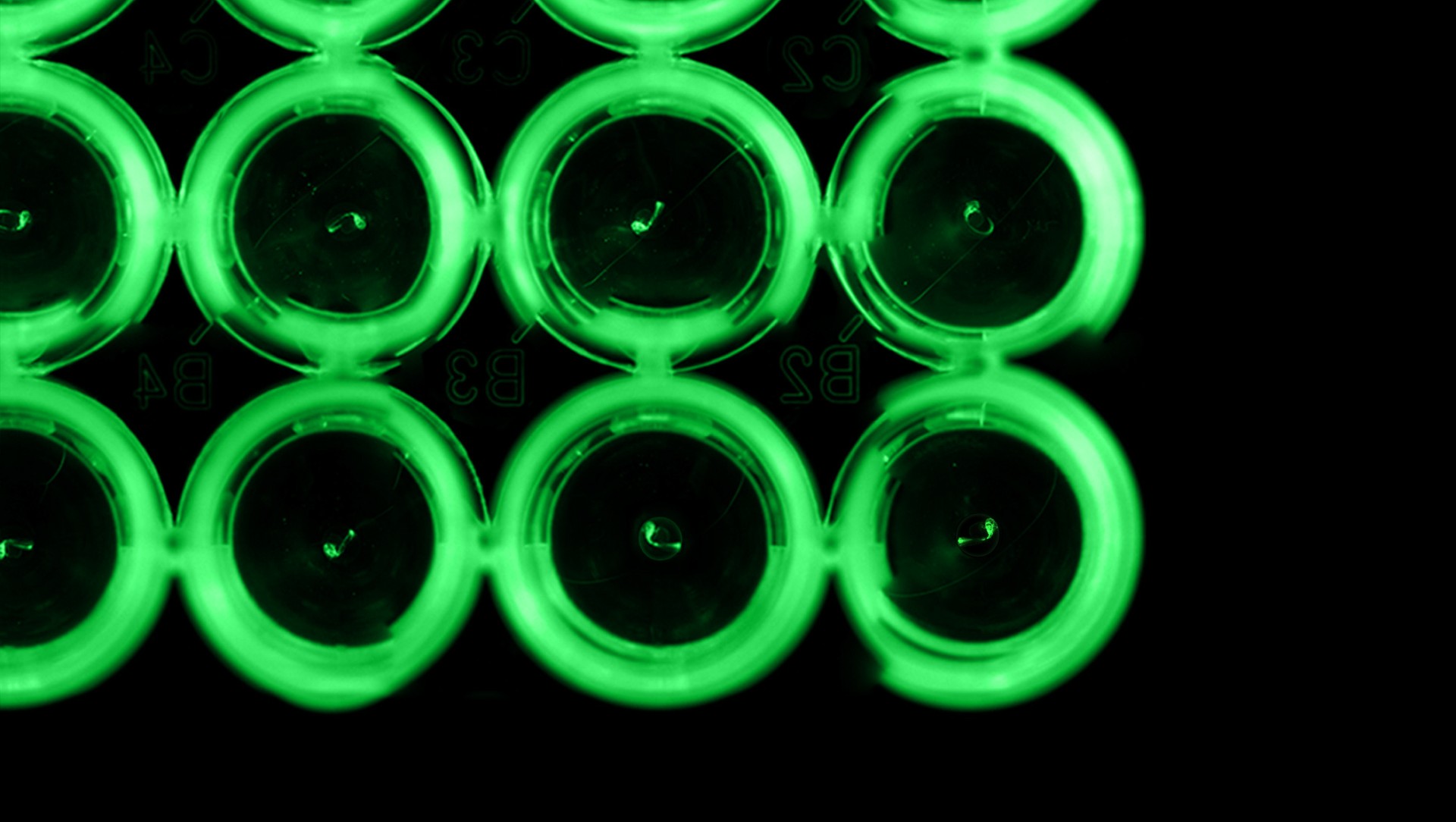
Embryo fluorescence heartbeat egg

OUR
PRODUCTS

Our computational microscopes are designed for todayā€™s researchers and built by a multi-disciplinary team of engineers, programmers, and scientists.
  • seconds to insight

    Our arrayed architecture allows for the capture of imaging data over an extremely wide field of view within a matter of seconds ā€” without sacrificing spatial resolution.
  • Preserve biological integrity

    With high-quality, synchronous data capture across entire well plates, set your imaging assays with confidence, knowing the results will be uniform and reliable.
  • sophisticated machine learning

    Our refined software provides users quick and easy solutions for tracking behavior, identifying key features, and quantifying activity.

the Kestrel

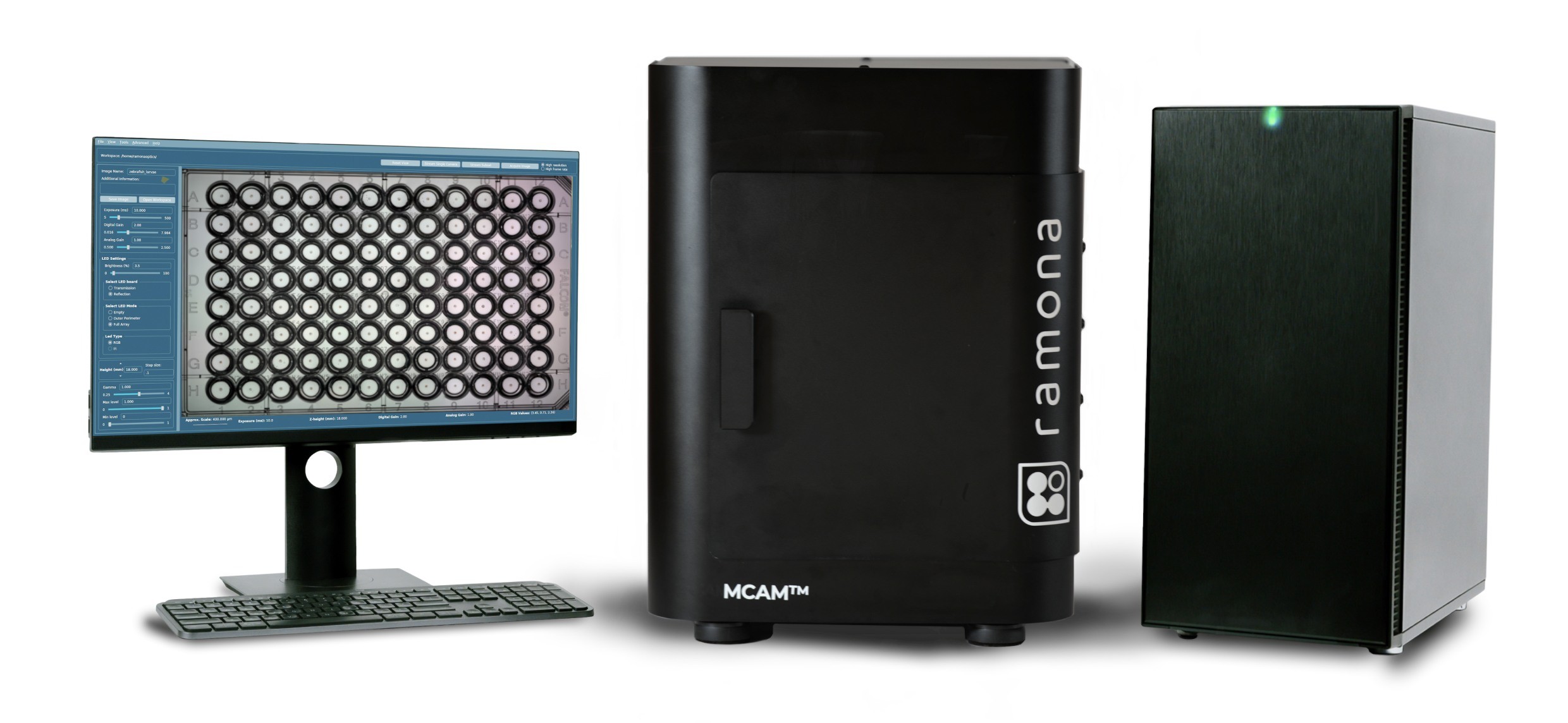
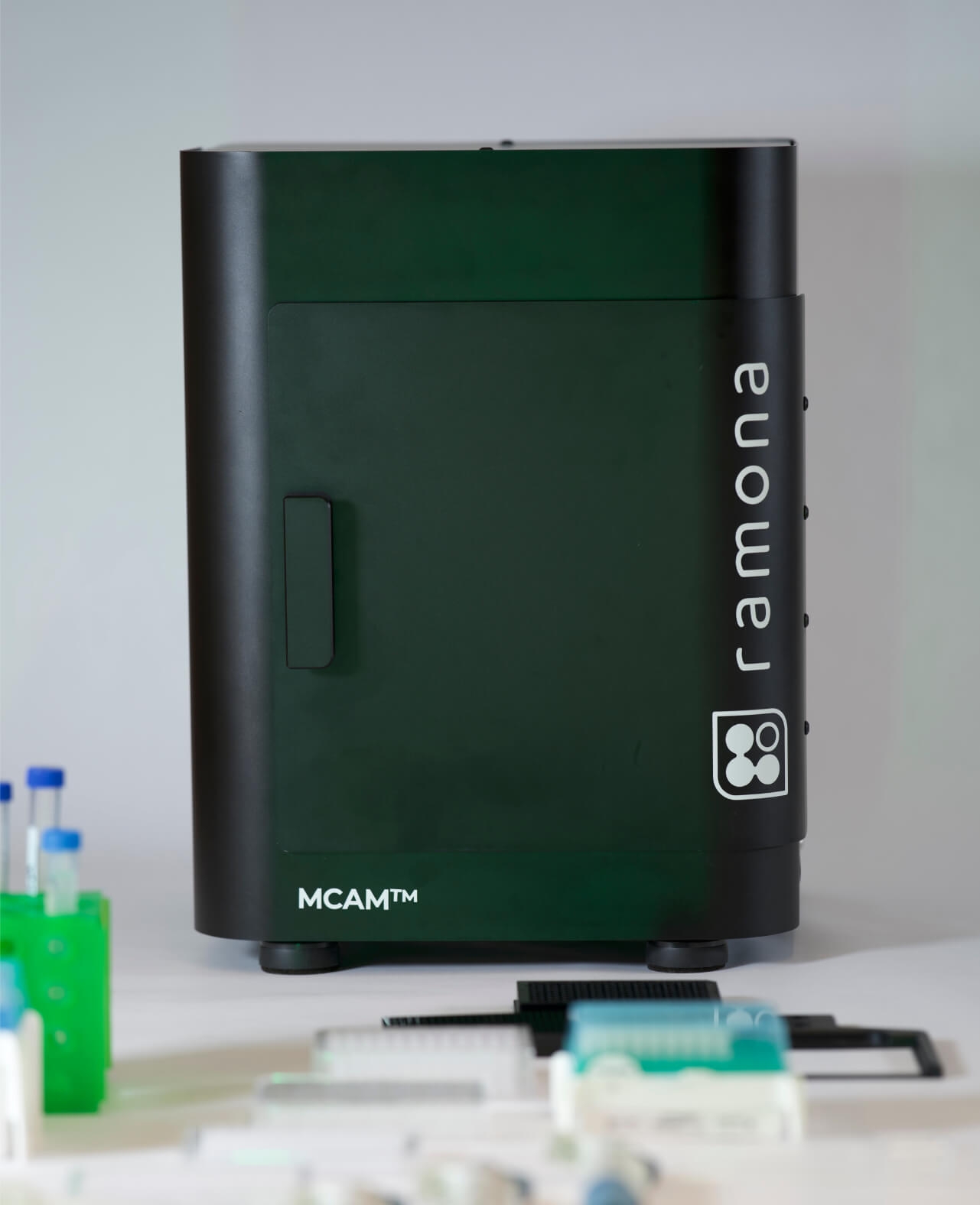
Our MCAMā„¢ Kestrel system is the high-throughput solution for researchers studying model organisms. Modular, fast, and easy to use, the MCAMā„¢ Kestrel is designed to shine in two main modes ā€” Behavior and Screening.
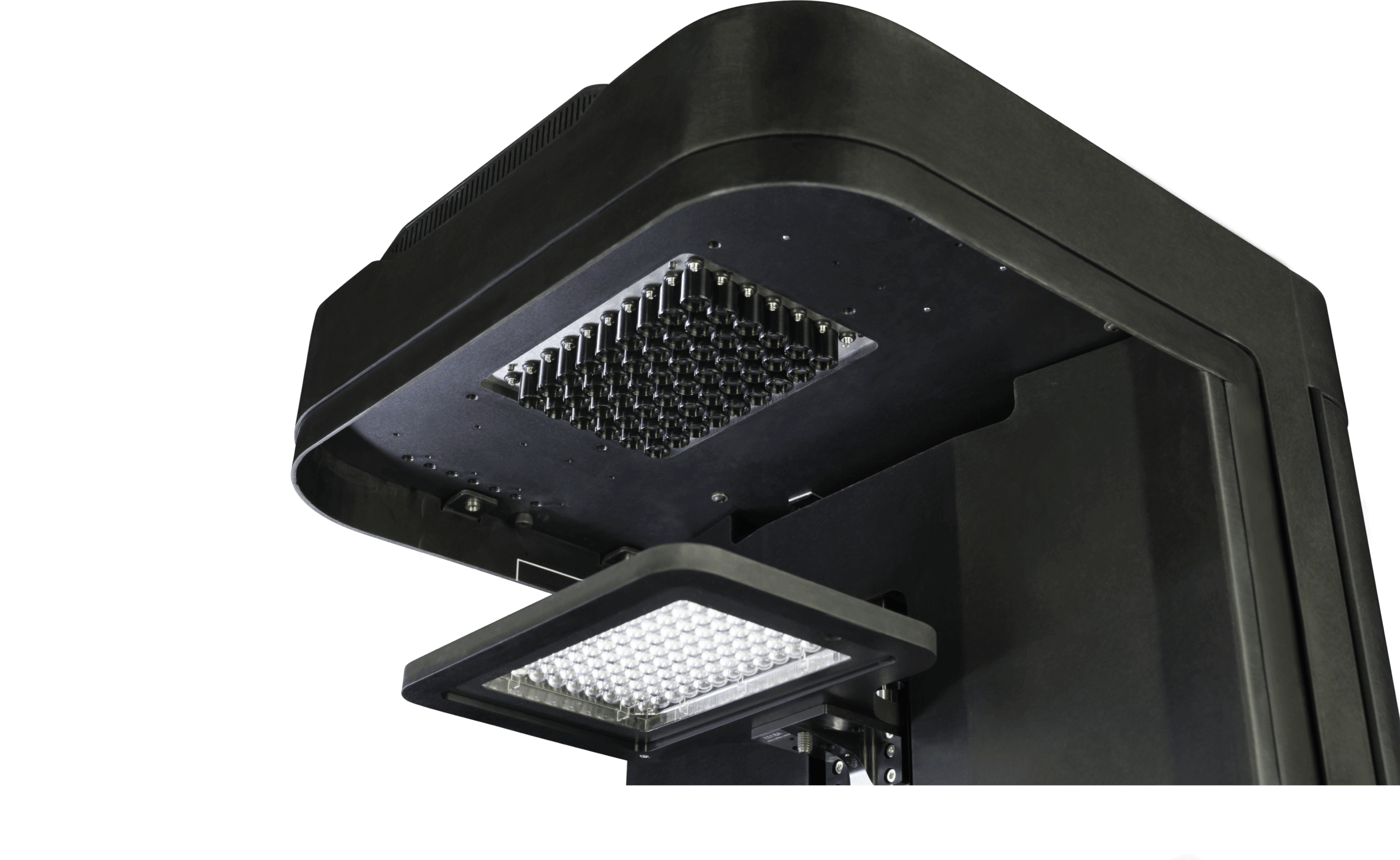
Our system includes a Multi-Camera Array Microscope (MCAMā„¢) and a powerful workstation for user operaiton and local data processing. Built from stainless steel and aluminum for repeated daily use, Ramona's MCAM puts the performance of a room full of microscopes on a standard lab bench.
behavior MODE
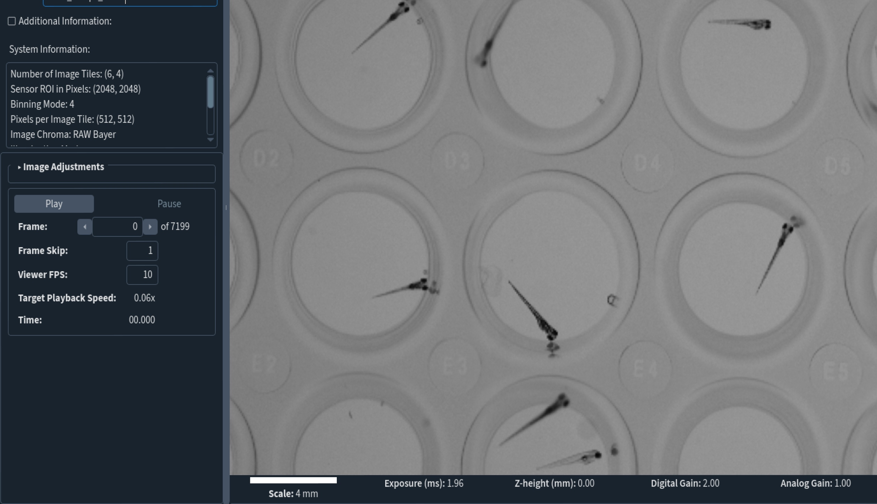
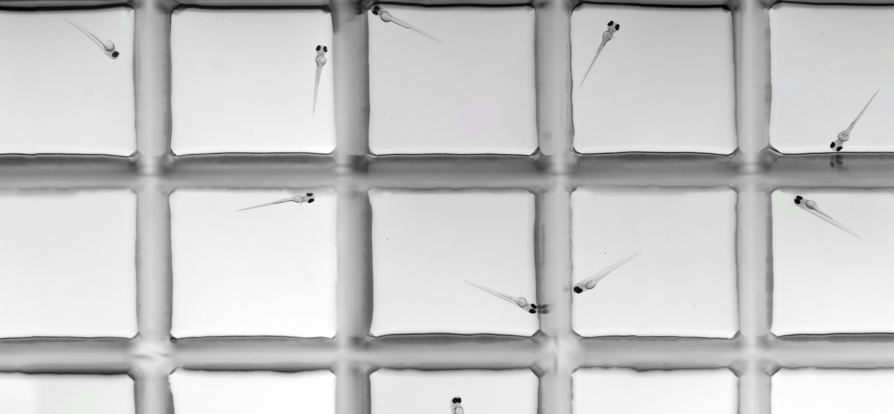
The MCAMā„¢ Kestrelā€™s Behavior Mode is designed to simultaneously observe all wells of a standard well plate with 24X higher spatial and temporal resolution than leading competitors. Rapidly quantify detailed locomotion kinematics and gross morphology with customized high-speed software to unlock new workflows for studying organism behavior at high throughput.
For more information, download supplementary materials below. For additional resources, contact us directly to schedule a virtual demo.
Technical SpecificationsProduct Sheet
Screening MODE
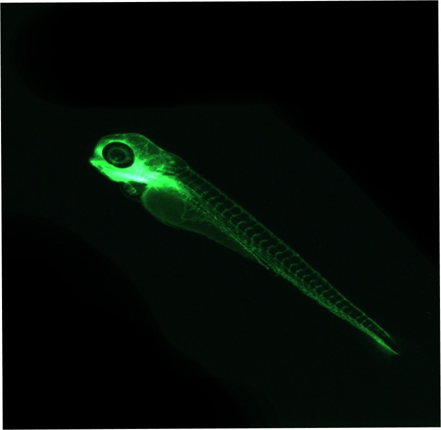
The MCAMā„¢ Kestrelā€™s Screening Mode is designed for higher-resolution, high-content microscopy. Our Screening Mode is equipped for parallelized functional fluorescence and brightfield imaging of small model organisms and quality control of organoid cultures. From fine-morphology and cardiac assessments, to fluorescence quantification and time lapse imaging, acquire multiple readouts throughout development in one integrated system.
For more information, download supplementary materials below. For additional resources, contact us directly to schedule a virtual demo.
Technical SpecificationsProduct Sheet

Ease
of use

Each multi-camera array microscope comes with a powerful storage system and an intuitive interface to easily design and implement protocols.
Our customers and development partners are advancing science every day.Our customers and development partners are advancing science every day, faster than ever.
  • National Institute of Health
  • Duke university
  • NC state university
  • UCSF School of University
  • Oregon state university
  • Mayo
  • West virginia
  • Harvard university
  • Tufts University
  • McLean
  • Northwestern University
  • AgBiome
  • Devcom
  • INRS
  • Cal
  • NSF logo
  • Corestemchemon
  • Universita Di Milano
  • UFZ

Behavior
Mode

System Features
  • High speed video

    In Behavior Mode, the MCAMā„¢ Kestrel captures subjects at up to 160fps, revealing fine movements invisible to the naked eye.Ā Distinguish between types of seizures, evaluate eye movements, and classify tail angle sequences during startle responses.
  • Skeletal tracking

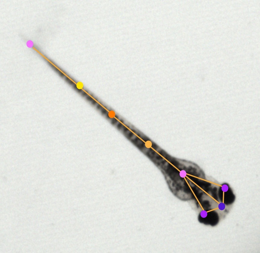
    Our 8-point skeletal overlay ignores debris and provides exact detail of fine, rapid movements.Ā  Use additional motion metrics for tail angle curvature analysis and startle responses.
  • Automated Morphology and Quality Control

    Use 8-point tracking to get information about the length and posture of your organisms. Ensure quality control of your stocks and compare behavioral and morphological differences across treatment groups.
  • Results
  • startle response
    See our MCAMā„¢ Kestrel administer a vibration stimulus and track zebrafish reactions with high-speed video.
  • Tail angle analysis
    View a larval Zebrafishā€™ tail curvature in detail, captured on high-speed video.
  • High-Throughput Tracking
    Learn how end-to-end behavioral experiments can be conducted and data analyzed over an entire 96-well plate using the MCAMā„¢ Kestrel.
  • Eye size + behavior
    See our MCAMā„¢ Kestrelā€™s eye angle tracking capacity over 96 wells.
User Feedback

The Tanguay Lab + Ramona

ā€œWe've been very impressed with the robustness of the MCAM platform and the quality of the data that it delivers. My lab uses the system everyday to streamline our well-plate based workflow - it has become a critical tool for us.ā€
Dr. Robyn L. Tanguay, PhD
Distinguished Professor in the Department of Environmental and Molecular Toxicology and Director of the Superfund Research Program, Oregon State University
Oregon state university
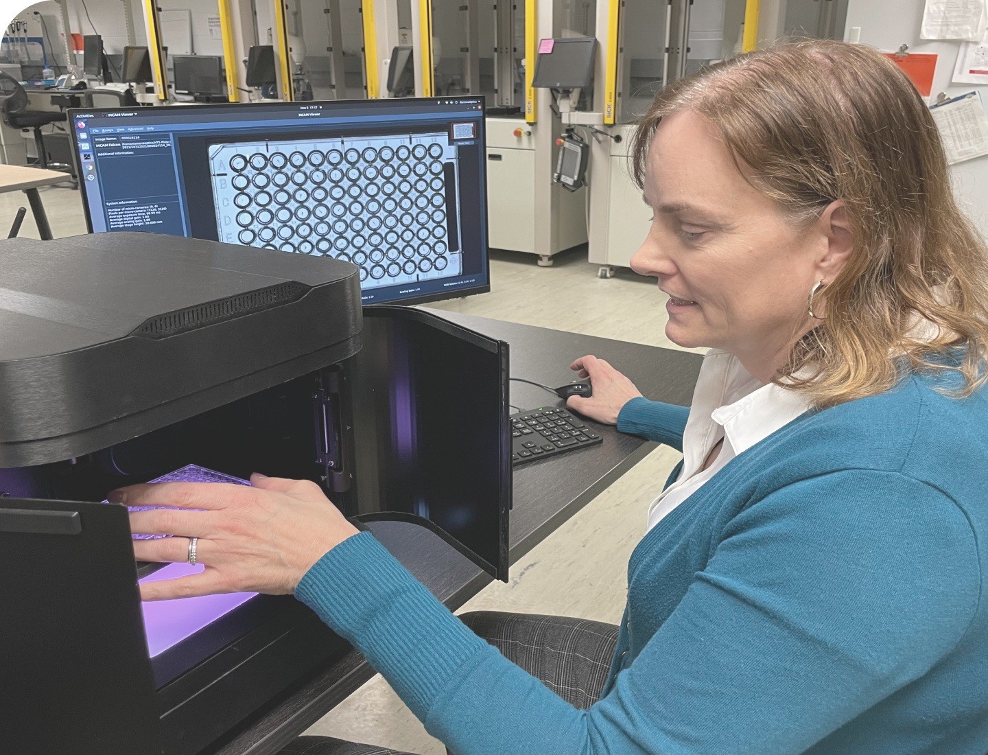
Under the direction of Dr. Tanguay, scientists at the world's largest zebrafish toxicology facility use Ramonaā€™s MCAM technology every day to quickly obtain reliable, quality data. At Ramona, we pride ourselves on building powerful tools that work day in and day out.
Screening
Mode

System Features
  • Fluorescence

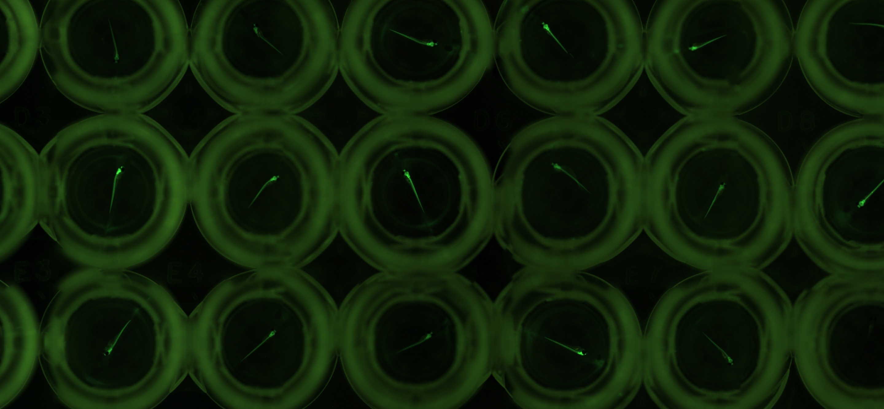
    In Screening Mode, the MCAMā„¢ Kestrel delivers comprehensive fluorescence imaging across entire well-plates. Built-in machine-learning analysis tools enable immediate data-driven decisions from uniform fluorescence results.
  • Timelapse

    Our time-lapse protocols allow for simultaneous snapshots across an entire well plate, eliminating time bias between samples and ensuring uniform data capture for studies on development and regeneration. Set-and-forget your assay once and capture uniform changes across all samples, from hours to days.
  • Integrated Analyses Pipelines

    Use our Morphology Classification, Neutrophil Quantification, and Heartbeat Quantification toolkits (and other bulk fluorescence analyses) to rapidly glean deeper insights from large datasets with little to no user input required.
  • Results
  • Neutrophils + Machine learning
    Read about the MCAMā„¢ Kestrelā€™s rapid, high-resolution algorithmic method to count fluorescent-labeledĀ cellsĀ in vivo.
  • Neutrophils on video
    Watch a quick video featuring data from a streamlined neutrophil quantification experiment in zebrafish larvae.
  • GFP Heartbeat
    Learn how the MCAMā„¢ Kestrel has been used to capture high-speed video of fluorescent cardiac activity synchronously over 96 wells.
  • Angiogenesis
    Read about how the MCAMā„¢ Kestrelā€™s automated machine learning can be used to quantify vasculature development in zebrafish larvae.
User Feedback

The Yoder Lab + Ramona

ā€œWe are excited about increasing throughput and statistical confidence for common assays in the zebrafish community by leveraging the high resolution data from the MCAM and automated image analysis software developed by Ramona.ā€
Dr. Jeffrey Yoder, PhD
Professor of Innate Immunology, North Carolina State University
NC state university
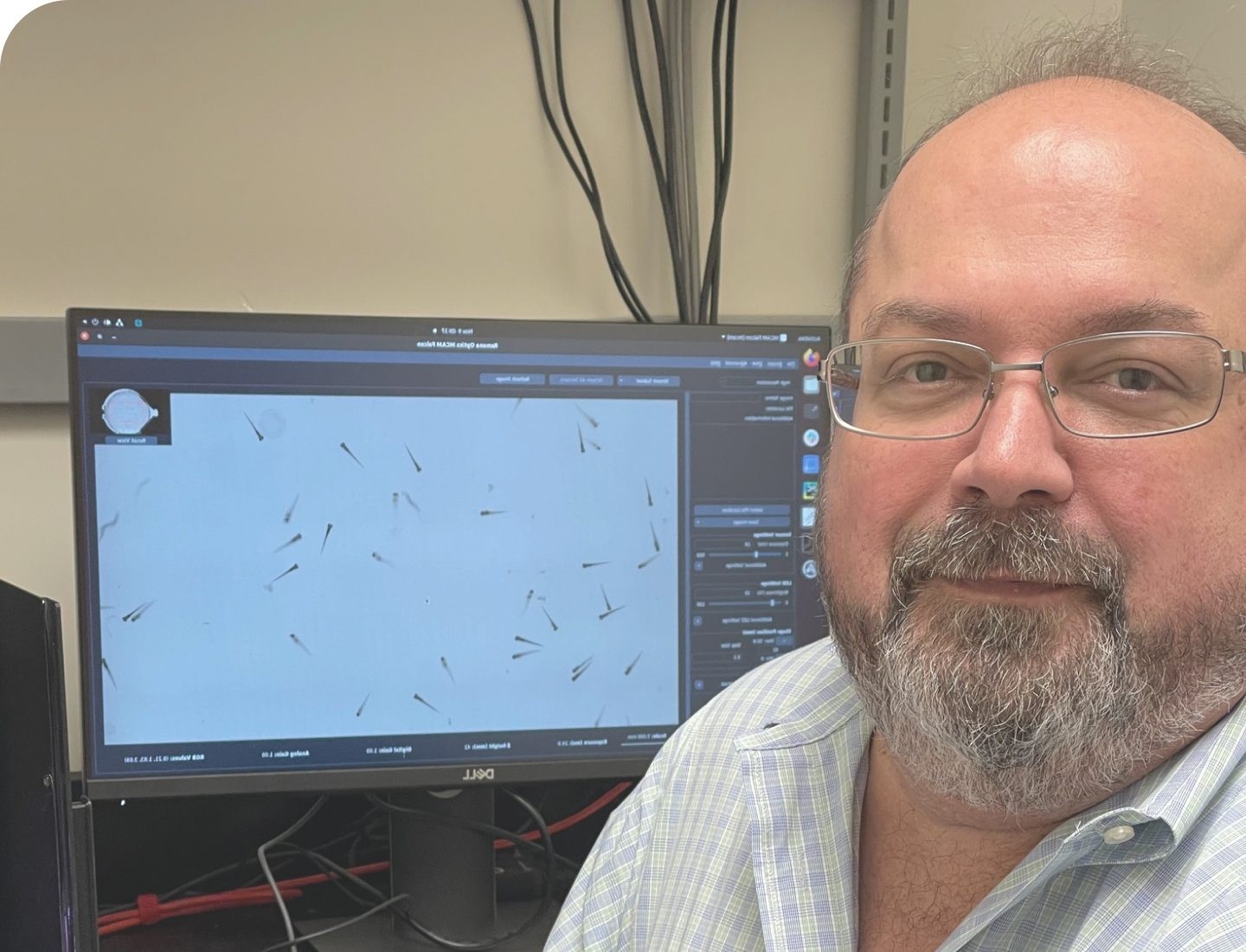
Ramona is working with the Yoder Lab to improve the efficiency and accuracy of chemical and genetic screens. With MCAM system automates the processing of high-resolution video to assess physiology, morphology, and imunnology of up to a hundred organisms simultanously. We are eager to help accelerate discovery by automating tedious measurements across rich and diverse datasets.
Learn more about how people are using the MCAM to unlock new workflows in the modern biology lab and beyond.
Visit Our Blog
To attach files, drag + drop here orĀ select files from your computer.